

Introduction
Wound management remains one of the perennial challenges in veterinary clinical practice. Whether resulting from trauma, surgical interventions, or chronic disease, open skin wounds in dogs demand therapeutic strategies that balance efficacy, safety, and practicality. While systemic medications have long been included in multimodal wound care protocols, the growing interest in topical pharmacotherapy has shifted the focus toward compounds capable of influencing local wound physiology without substantial systemic absorption.
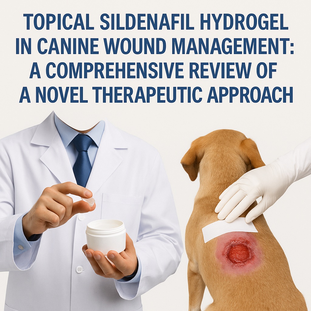
Among such agents, sildenafil citrate—widely known for its vasodilatory properties through inhibition of phosphodiesterase-5 (PDE5)—has attracted particular attention. Originally developed for cardiovascular applications and popularized for the management of erectile dysfunction, sildenafil possesses additional biological activities that extend well beyond smooth muscle relaxation. In fact, literature over the past two decades has documented its effects on angiogenesis, nitric oxide metabolism, oxidative stress reduction, and microcirculatory enhancement—all of which are intimately interconnected with wound repair processes. Several studies in rodents and experimental models have demonstrated improved wound perfusion, flap viability, epithelialization, and granulation tissue formation after sildenafil exposure. Nevertheless, controlled studies evaluating topical sildenafil in dogs have been conspicuously absent.
The research summarized in this article fills that gap by presenting a meticulously designed investigation of a novel sildenafil citrate hydrogel, formulated for direct application to open wounds in dogs. Drawing on the detailed experimental findings from the original report , the following discussion interprets its scientific relevance, evaluates its therapeutic potential, and reflects on its implications for future veterinary wound care.
The Rationale Behind Topical Sildenafil: Biological Foundations
The choice to explore sildenafil as a topical wound agent is grounded in its multifaceted biochemical actions. At the cellular level, sildenafil enhances nitric oxide (NO) signaling by inhibiting PDE5, slowing the breakdown of cyclic guanosine monophosphate (cGMP). This prolongs vasodilation and improves local blood flow—an effect that is particularly important in wounded, ischemic, or poorly perfused tissues.
NO itself plays several essential roles in wound repair:
- It modulates platelet activity, reducing excessive clot rigidity and promoting physiologic hemostasis.
- It contributes to microbial defense, offering antimicrobial effects within the wound bed.
- It reduces oxidative stress by neutralizing reactive oxygen species produced during injury.
- It fosters angiogenesis, fibroblast activity, and cellular proliferation—all critical to tissue regeneration.
Numerous rodent studies have described accelerated healing with topical sildenafil application, including improved flap survival, enhanced granulation tissue, and strengthened collagen architecture. Interestingly, one study cited in the original publication demonstrates that topical sildenafil may be more effective than oral administration for select wound types. Such evidence lays a solid physiological foundation for exploring a topical formulation in canine models.
However, translating rodent research to dogs requires careful consideration, as canine dermal structure, inflammatory responses, and healing timelines differ significantly from those of smaller laboratory animals. The absence of prior controlled topical sildenafil studies in dogs made this investigation particularly timely and clinically relevant.
Formulation of a Novel Sildenafil Hydrogel: Chemical and Mechanical Characteristics
Developing a topical wound product involves far more than simply dissolving a drug in a base. Stability, spreadability, viscosity, uniformity, and release kinetics must all be tailored to ensure safe and effective delivery. The research team conducted extensive solubility testing and selected poloxamer 407 hydrogel as the primary vehicle, supplemented with propylene glycol and N-methyl-2-pyrrolidone as pharmaceutical solvents.
The resulting formulations contained 0%, 5%, or 10% sildenafil citrate and demonstrated several key characteristics:
- Homogeneity – Uniform distribution of sildenafil throughout the gel was confirmed with high-performance liquid chromatography.
- Shear-thinning viscosity – All hydrogels became less viscous under shear force, a desirable feature that allows easy application while maintaining structure once in place.
- Controlled drug release – In vitro testing revealed that more than 90% of sildenafil was released within 8 hours, reflecting a sustained yet predictable release profile well suited to once-daily bandage changes.
- Acidic pH – The gels exhibited pH values between 3.9 and 5.3. This slight acidity may inherently support wound healing by enhancing antimicrobial activity and stimulating granulation tissue.
This formulation strategy underscores the complexities of pharmaceutical design and helps explain why the hydrogel—especially in sildenafil-containing concentrations—performed as it did in vivo.
Study Design: Controlled Evaluation in a Canine Wound Model
To test the hydrogel’s clinical utility, the authors used a standardized, reproducible model: full-thickness 3 × 3 cm excision wounds created along the dorsum of six purpose-bred adult Beagles. Each dog received four wounds that were randomly assigned to one of four treatments:
- C (Control): nonadherent bandage only
- G: base hydrogel without sildenafil
- 5S: hydrogel with 5% sildenafil
- 10S: hydrogel with 10% sildenafil
Daily wound assessments, high-resolution imaging, and precise planimetry allowed quantification of wound size, contraction rates, and epithelialization. Histopathology samples were collected across multiple time points to characterize inflammation, fibroblast proliferation, collagen deposition, and neovascularization. Furthermore, plasma sildenafil levels were monitored to evaluate systemic absorption and safety.
This rigorous, blinded, internally controlled design lends strong credibility to the study’s findings and ensures that observed effects were attributable to the hydrogel composition rather than individual or environmental variability.
Early Granulation Tissue Formation: The Most Striking Outcome
One of the clearest findings was the accelerated formation of granulation tissue in wounds treated with sildenafil hydrogels. The mean time to first granulation tissue appearance was:
- 2.8 ± 0.8 days for both 5% and 10% sildenafil
- 5.2 ± 0.4 days for the control
- 6.3 ± 1.4 days for the base hydrogel
In other words, sildenafil shortened the onset of granulation by approximately 50% compared to a standard nonadherent dressing and by more than half compared to the hydrogel base alone.
Granulation tissue is essential for successful secondary closure, grafting, and natural wound contraction. An earlier granulation response may substantially alter the clinician’s ability to close wounds surgically or guide them toward favorable outcomes.
Interestingly—and somewhat ironically—the base hydrogel alone delayed granulation tissue formation and slowed contraction. This demonstrates that a wound vehicle is not always inert, and underscores the importance of formulation research rather than assuming that any “gel” will do.
Wound Contraction and Epithelialization: Dose-Dependent Trends
The presence of sildenafil improved wound contraction rates relative to the base hydrogel. Wounds treated with the G formulation contracted 10–14% less over the study period than wounds in the other groups, confirming that sildenafil largely counteracted the contraction-slowing effects of the base formulation.
Between the 5% and 10% sildenafil groups, the differences were subtler:
- 10S wounds were slightly smaller than 5S wounds by day 21 (an average difference of 0.7 cm²).
- No significant differences were observed between the active sildenafil groups and the simple nonadherent dressing in overall contraction or epithelialization.
Nevertheless, the timing of these events likely holds deeper clinical meaning. Sildenafil’s most pronounced influence occurred early, during the inflammatory and early proliferative phases—suggesting that it may serve best as a short-term adjunct to jump-start delayed healing rather than a long-term treatment through all stages of repair.
Inflammation and Reparative Histology: A Balancing Act
Histopathologic assessments revealed an intriguing pattern:
- Higher early acute inflammation (day 3) in sildenafil-treated wounds, possibly related to increased perfusion and immune cell recruitment.
- Higher reparative scores (day 7), consistent with accelerated granulation.
- Higher late inflammation (day 21) in sildenafil groups, which may suggest lingering immune activity or delayed resolution of the inflammatory phase.
Neovascularization—a cornerstone of healthy wound repair—was significantly greater in the 10S group on days 7 and 14, supporting the known angiogenic effects of sildenafil. More robust vascularization often correlates with better tissue oxygenation, improved fibroblast function, and more efficient collagen deposition.
The presence of slightly elevated late inflammation in sildenafil groups does not necessarily denote impaired healing—indeed, inflammation and proliferation are tightly interdependent—but it suggests that these formulations might benefit from early-phase, limited-duration use rather than prolonged continuous therapy.
Systemic Absorption: A Safety Perspective
One of the most important clinical considerations in topical therapy is whether systemic absorption may lead to unintended drug exposure. In this study, most dogs had no detectable plasma sildenafil throughout the experiment. However, a few dogs exhibited low-to-moderate plasma concentrations after two to three weeks of daily topical application.
These levels remained within known safe ranges for canine sildenafil therapy and were far below the concentrations achieved through standard oral dosing regimens. No adverse effects were reported.
Nevertheless, clinicians should remain mindful of the potential for systemic absorption, especially in:
- Large wounds
- Highly vascularized wound beds
- Patients receiving concurrent vasodilators
- Dogs with cardiopulmonary disease
Further studies would help clarify dosing frequency, ideal treatment duration, and long-term systemic safety.
Clinical Significance and Practical Implications
The findings of this study suggest that topical sildenafil hydrogel may be an effective tool for stimulating early granulation tissue, potentially benefiting clinical scenarios such as:
- Traumatic degloving injuries
- Surgical wounds healing by second intention
- Chronic non-granulating ulcerations
- Wounds requiring later grafting or reconstructive surgery
However, the data also caution against assuming a simple linear benefit. The base hydrogel delayed contraction and epithelialization, and sildenafil’s advantages were most pronounced during early healing rather than throughout all phases. In practice, topical sildenafil may best serve as a phase-specific intervention—something akin to a starter boost for stalled or ischemic wounds.
Before widespread clinical adoption, further research is warranted to refine the vehicle formulation, optimize pH balance, evaluate alternative hydrogels, and determine ideal treatment schedules. Larger clinical trials in diverse canine populations (beyond healthy young Beagles with controlled surgical wounds) would also strengthen the evidence base.
Conclusion
This investigation into a novel sildenafil citrate hydrogel provides compelling evidence that topical sildenafil can meaningfully influence early wound healing in dogs. By accelerating granulation tissue formation and enhancing early reparative processes, sildenafil demonstrates promising potential as a targeted adjunctive therapy in veterinary wound management.
At the same time, the study reminds clinicians that formulation matters, timing matters, and systemic absorption—though limited—is not negligible. As with many pharmacologic innovations, topical sildenafil represents an exciting beginning rather than an endpoint. Continued refinement and clinical evaluation are likely to determine its ultimate place in the veterinary wound care toolkit.
FAQ: Frequently Asked Questions
1. Can topical sildenafil replace standard wound care for dogs?
No. Based on current evidence, topical sildenafil should be viewed as an adjunct, not a replacement. While it accelerates early granulation tissue formation, it does not outperform standard dressings in contraction or epithelialization. Traditional wound hygiene, protection, and decontamination remain essential components of care.
2. Is topical sildenafil safe for long-term daily use?
Short-term use appears safe, with minimal systemic absorption and no reported adverse effects. However, low-level systemic absorption was detected in some dogs after 14–21 days. Until more data are available, prolonged continuous application should be used cautiously, especially in dogs with cardiovascular disease.
3. Which wounds are most likely to benefit from topical sildenafil?
Wounds lacking early granulation tissue—such as chronic ulcers, ischemic wounds, or large traumatic lesions—may benefit the most. Topical sildenafil is particularly promising for situations in which early granulation is crucial for later surgical closure.
