

Introduction
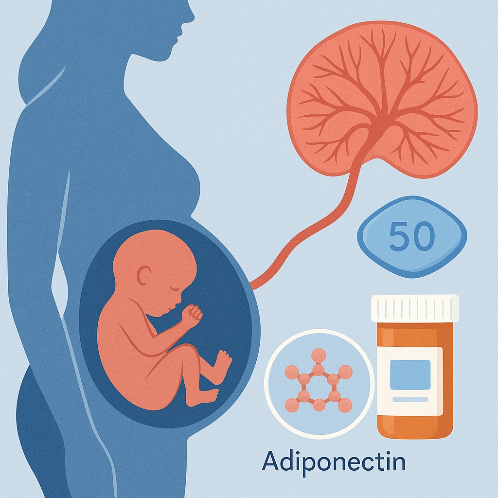
The concept of using sildenafil—a drug known worldwide for its role in male erectile dysfunction—to modulate fetal growth might sound like the opening line of an academic satire. Yet modern reproductive endocrinology thrives exactly at such unexpected intersections. As our understanding of placental biology deepens, it becomes increasingly clear that the placenta is not a passive conduit but a metabolically rich, hormonally responsive organ profoundly influenced by maternal endocrine and vascular signals. This insight becomes crucial when considering gestational environments altered by hyperandrogenism, a hallmark of polycystic ovary syndrome (PCOS), one of the most prevalent endocrine disorders affecting reproductive-aged women.
The article under analysis investigates a highly specific but clinically relevant phenomenon: the impact of sildenafil on fetal weight and placental adiponectin expression in pregnant rats rendered glucose intolerant through gestational testosterone exposure. This model mimics several essential features of PCOS pregnancies: hyperandrogenemia, impaired glucose tolerance, disrupted placental signaling, and reduced fetal growth. On this scientific stage, sildenafil is introduced not as a sexual medicine agent but as a vasodilator capable of modifying placental perfusion and metabolic signaling.
In the following sections, we dissect the mechanistic rationale, experimental design, and outcomes of this study, connecting the authors’ findings with broader implications for maternal–fetal medicine and metabolic endocrinology. While the journey may traverse biochemical pathways and hormonal loops, the conclusion is elegant in its simplicity: sildenafil, through its vascular and metabolic modulatory effects, partially rescues placental dysfunction and improves fetal growth in a compromised intrauterine environment.
Understanding the Model: Testosterone-Induced Gestational Glucose Intolerance
To appreciate the significance of the findings, one must first understand the biological model used in the study. The authors employed pregnant rats exposed to testosterone throughout gestation to induce glucose intolerance—an approach with strong parallels to the metabolic impairments observed in PCOS pregnancies. The PDF details that maternal hyperandrogenemia disrupts insulin signaling pathways, increases systemic inflammation, and negatively affects placental nutrient transport, ultimately restricting fetal growth.
Indeed, the glucose tolerance tests shown in the figures (pages 4–5) demonstrate higher glucose excursions in testosterone-treated pregnant rats compared to controls, confirming metabolic impairment. The hyperandrogenic intrauterine environment not only affects maternal physiology but also disrupts placental endocrine signaling. One key player in this disrupted network is adiponectin, a hormone known for its insulin-sensitizing, anti-inflammatory, and pro-angiogenic properties.
Maternal adiponectin typically declines in pathological pregnancies associated with insulin resistance, while placental adiponectin expression is altered in response to hormonal and vascular stress. In testosterone-exposed pregnancies, the placenta shows reduced adiponectin protein levels (page 7), along with morphological evidence of impaired development and reduced fetal weight.
This model is therefore well-suited to study interventions that target placental perfusion or metabolic signaling—and sildenafil, with its dual influence on vascular tone and endothelial nitric oxide pathways, emerges as a logical candidate.
Why Sildenafil? Mechanistic Rationale Beyond Vasodilation
Sildenafil’s biological function extends well beyond its well-known pharmacological niche. By inhibiting phosphodiesterase-5 (PDE5), sildenafil increases intracellular cGMP levels, enhancing smooth muscle relaxation and improving tissue perfusion. In the reproductive field, PDE5 is expressed in the uterus and placenta, and sildenafil has been shown to improve uteroplacental blood flow in several animal models.
But there is more. The placenta is not simply a vascular organ—it is a hub of metabolic signaling. Increased cGMP influences angiogenesis, trophoblast differentiation, and nutrient transporter expression, all of which can modify fetal growth trajectories.
In the current study, sildenafil is not used to increase maternal systemic glucose tolerance—indeed, the researchers show that sildenafil did not reverse glucose intolerance in testosterone-treated rats (page 6). Instead, its influence appears more targeted: improving placental microenvironment, restoring hormones essential for nutrient regulation, and ultimately enhancing fetal weight.
Thus, sildenafil’s role in this study is not to “treat diabetes” in pregnancy, but to modulate the downstream consequences of gestational hyperandrogenemia at the placental level.
Effects on Maternal Glucose Tolerance: What the Study Reveals
An important finding—presented clearly in glucose tolerance curves (page 5)—is that sildenafil did not normalize glucose metabolism in testosterone-exposed pregnant rats. Despite known vasodilatory activity, sildenafil did not significantly change maternal glucose levels at any measured time point.
This finding underscores a crucial point:
Sildenafil’s beneficial effects in this model occur independently of systemic maternal glucose homeostasis.
By ruling out maternal glycemic improvement as a confounding variable, the authors strengthen the argument that sildenafil exerts its effects directly (or at least primarily) on the placenta and fetus. This distinction is essential when interpreting the next stage of results: the biological rescue of fetal weight.
Impact on Fetal Weight: Sildenafil as a Placental Modulator
According to Table 1 and associated graphs (page 6), gestational testosterone exposure significantly reduced fetal weight compared to controls, confirming the negative impact of hyperandrogenic pregnancy on fetal development.
When sildenafil was administered concomitantly with testosterone:
- Fetal weight increased significantly, approaching values seen in healthy controls.
- Placental weight remained altered, suggesting that sildenafil did not simply enlarge or “bulk up” the placenta.
- The fetal-to-placental ratio improved, indicating more efficient placental function.
This is a key point: sildenafil does not cause pathological placental enlargement but appears to restore functional capacity. The fetal weight gain is therefore not an artifact of placental swelling but a result of improved nutrient transfer and metabolic signaling.
It’s noteworthy that fetal rescue occurred despite no change in maternal glucose handling. This strongly supports the hypothesis that sildenafil’s effect is mediated through the placenta.
Placental Adiponectin: A Central Mediator in Fetal Growth
One of the most scientifically intriguing results in the study is the impact of sildenafil on placental adiponectin. Western blot analyses (page 7) demonstrate that:
- Gestational testosterone reduces placental adiponectin protein expression.
- Sildenafil significantly restores adiponectin levels in testosterone-exposed placentas.
- mRNA levels of adiponectin remain comparatively unchanged, indicating post-transcriptional regulation.
This nuance is compelling. Sildenafil did not increase adiponectin gene transcription—rather, it appears to stabilize adiponectin protein levels or influence its processing or degradation. The precise mechanism remains unclear, but several biological pathways are possible:
- cGMP may inhibit proteasomal degradation of adiponectin.
- Enhanced placental perfusion may reduce oxidative stress, protecting adiponectin protein.
- Improved endothelial function may modulate placental adiponectin secretion or recycling.
Whatever the mechanism, restoring adiponectin is physiologically meaningful. Adiponectin enhances insulin sensitivity, increases fatty acid oxidation, and regulates placental nutrient transfer—all factors essential for healthy fetal growth.
The correlation between adiponectin restoration and increased fetal weight supports the idea that placental endocrine correction is a major pathway through which sildenafil improves fetal outcomes.
Morphological and Histological Findings: Beyond Numbers
Histological images presented in the study (page 8) provide visual evidence of placental alterations. In testosterone-exposed rats, placentas show:
- reduced labyrinthine zone density
- increased trophoblast disorganization
- signs of impaired vascular architecture
Sildenafil-treated placentas, in contrast, show:
- better vascular arrangement
- more organized trophoblastic layers
- improved labyrinth structure
These findings align with sildenafil’s known angiogenic and vasodilatory effects. Improved labyrinthine structure suggests enhanced maternal–fetal exchange, which likely contributes to the increased fetal weight.
Interestingly, these improvements occur without full normalization of placental morphology, reflecting a partial rather than complete rescue. This nuance indicates that sildenafil ameliorates but does not reverse the adverse effects of gestational testosterone—important for managing expectations in translational research.
Mechanistic Pathways: How Does Sildenafil Restore Placental Function?
Based on the PDF’s data and the current literature, several mechanistic pathways may explain how sildenafil improves fetal growth in this context.
1. Enhanced Uteroplacental Blood Flow
Sildenafil increases cGMP in vascular endothelial cells, improving:
- vasodilation
- perfusion
- oxygenation
Better perfusion enhances nutrient delivery, which is particularly critical in compromised pregnancies.
2. Reduction of Placental Oxidative Stress
Hyperandrogenic environments increase oxidative stress. Sildenafil has been shown in other studies to improve antioxidant enzyme activity, potentially protecting placental proteins such as adiponectin.
3. Stabilization of Adiponectin Protein
Not through gene transcription, but via modulation of post-translational pathways—an insight specifically highlighted by the mismatch between protein and mRNA levels.
4. Improved Trophoblast Function
Optimal cGMP signaling supports trophoblast invasion and differentiation, two processes disrupted by gestational androgen excess.
5. Normalization of Placental Endocrine Crosstalk
Restoring adiponectin creates downstream benefits for placental glucose transport, lipid metabolism, and nutrient allocation.
In short, sildenafil appears to “unlock” dysfunctional placental signaling, improving its efficiency without altering maternal glucose metabolism.
Limitations and Interpretative Considerations
The authors appropriately acknowledge limitations:
- This is an animal model; human extrapolation must be cautious.
- Testosterone-induced glucose intolerance may not capture the full complexity of PCOS pregnancies.
- Sildenafil’s pharmacokinetics differ between rats and humans.
- The study does not explore long-term postnatal outcomes, an essential consideration in fetal programming research.
- It remains unclear whether adiponectin restoration is mechanistically central or simply correlated with improved placental function.
These limitations should not diminish the relevance of the study; rather, they highlight areas for future research, including human clinical trials focused on placental endocrine function rather than maternal glycemia.
Conclusion
This study offers an elegant demonstration of how sildenafil modulates the placental environment in a metabolically compromised pregnancy. By restoring placental adiponectin protein levels, improving trophoblast structure, and enhancing fetal weight, sildenafil shows promise as a therapeutic agent aimed at the placental–fetal interface rather than the maternal metabolic system.
The implications reach beyond reproductive endocrinology. They suggest that placental dysfunction in hyperandrogenic pregnancies may be amenable to targeted pharmacological rescue, even when maternal endocrine abnormalities persist. While sildenafil is unlikely to become a mainstream therapy for at-risk pregnancies without further research, this study lays essential groundwork and reinforces a powerful insight:
sometimes supporting the placenta is enough to change the course of fetal development.
FAQ
1. Does sildenafil treat maternal glucose intolerance in this model?
No. The study shows clearly that sildenafil does not correct glucose intolerance in testosterone-exposed rats. Its beneficial effects occur at the placental level, not through improving maternal metabolism.
2. Why is placental adiponectin important?
Placental adiponectin regulates nutrient transport, insulin sensitivity, inflammation, and trophoblast function. Reduced adiponectin is linked to fetal growth restriction. Sildenafil restores adiponectin protein levels, improving placental efficiency.
3. Could sildenafil be used in human pregnancies?
Although promising, current evidence is insufficient for clinical use. Human placental function is more complex, and long-term fetal safety must be established. This study provides a mechanistic foundation but not a clinical recommendation.
