

Introduction: The Persistent Puzzle of Fetal Growth Restriction
Intrauterine growth restriction (IUGR) remains one of the most pressing challenges in obstetric medicine. Globally, between 5% and 55% of pregnancies are affected depending on region and maternal risk factors. The consequences are profound: babies born small for gestational age carry an increased risk of perinatal mortality, impaired neurodevelopment, and lifelong cardiometabolic disease. Understanding its origins, and more importantly, finding effective therapeutic strategies, has become a major scientific pursuit.
Traditional explanations of IUGR have centered on maternal malnutrition, placental insufficiency, and exposure to toxins such as nicotine. However, endocrinology has added a new layer of complexity. It is now recognized that elevated maternal androgens, particularly testosterone, can impair placental function and restrict fetal growth. This phenomenon is not merely theoretical: women with conditions such as polycystic ovary syndrome (PCOS) or preeclampsia often demonstrate both high circulating testosterone and an increased frequency of growth-restricted neonates.
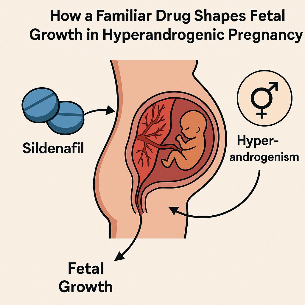
Amid these discoveries, researchers have begun to question whether drugs known for vascular modulation and metabolic effects might protect the placenta and fetus from the deleterious influence of androgens. One such candidate is sildenafil, a phosphodiesterase type 5 inhibitor (PDE5i) best known for its use in male erectile dysfunction. Its ability to enhance nitric oxide-mediated vasodilation has prompted investigations into whether it can improve uteroplacental blood flow and correct fetal growth abnormalities. The study at hand, performed in pregnant rats exposed to excess testosterone, provides fresh insights into how sildenafil acts at the maternal-fetal interface.
Hyperandrogenism in Pregnancy: An Unwanted Guest
Pregnancy is typically characterized by finely tuned hormonal orchestration. Unfortunately, when testosterone rises excessively, the delicate balance is disrupted. Evidence from both human and animal studies shows that hyperandrogenic pregnancies are linked to impaired glucose regulation, abnormal lipid handling, and oxidative stress across multiple maternal tissues. These disturbances do not spare the placenta—the critical interface responsible for nutrient and oxygen exchange between mother and fetus.
Animal experiments have consistently demonstrated the negative impact of maternal testosterone exposure. In sheep, administration of testosterone reduces the birth weight of both male and female offspring. In rats, it induces metabolic disturbances ranging from hepatic lipid accumulation to impaired antioxidant defenses in the maternal heart. Translating these findings to humans, the parallels are striking. Women with PCOS exhibit higher androgen levels, and their infants are more frequently small for gestational age. Similarly, elevated testosterone has been documented in preeclampsia, a condition notorious for poor fetal outcomes.
The precise mechanisms through which testosterone exerts its growth-restrictive effects are still being elucidated. Oxidative stress, inflammation, and mitochondrial dysfunction appear central. Excess androgens increase the generation of reactive oxygen species, depress antioxidant defenses, and trigger redox imbalance. The placenta, already a high-energy organ, becomes metabolically inefficient under these conditions, failing to adequately nourish the fetus. This sets the stage for IUGR—a condition not simply of restricted blood supply, but of defective placental biochemistry.
The Placenta as a Battlefield of Oxidative Stress
The placenta is not a passive structure; it is metabolically dynamic and highly sensitive to redox fluctuations. When oxidative stress predominates, several damaging cascades are unleashed. Protein malfunction impairs transport of glucose and amino acids. Lipid peroxidation damages cellular membranes, while mitochondrial dysfunction hampers energy production. These combined effects compromise placental efficiency, leading to smaller, undernourished fetuses.
One of the body’s key defenses against oxidative stress is the nuclear factor erythroid 2-related factor 2 (Nrf2) pathway. When activated, Nrf2 induces the transcription of numerous antioxidant enzymes including superoxide dismutase, catalase, and glutathione-related proteins. In healthy pregnancies, Nrf2 helps preserve placental function despite the inevitable oxidative burden of gestation. However, in hyperandrogenic pregnancies, Nrf2 signaling appears blunted. Studies have suggested that testosterone interferes with Nrf2 activation, leaving the placenta vulnerable to oxidative assault.
Alongside Nrf2, adiponectin—an anti-inflammatory adipokine expressed in the placenta—emerges as another key player. Adiponectin has complex roles: while some studies suggest it may restrict nutrient transfer, others consistently show that higher placental adiponectin correlates with greater fetal weight. Importantly, adiponectin is known to modulate oxidative pathways, including interactions with Nrf2, thereby bridging metabolic regulation and antioxidant defense. This dual role makes it a molecule of great interest in IUGR research.
Sildenafil: More Than a Vasodilator
Although sildenafil is universally recognized for treating erectile dysfunction, its pharmacological profile extends well beyond the corpus cavernosum. By inhibiting PDE5, the enzyme responsible for degrading cyclic guanosine monophosphate (cGMP), sildenafil prolongs nitric oxide signaling, leading to potent vasodilation. This mechanism has already found therapeutic application in pulmonary hypertension and certain high-altitude conditions.
But sildenafil’s repertoire does not stop at vascular smooth muscle. Studies show it can protect mitochondrial function, enhance antioxidant defenses, and modulate cellular signaling pathways such as PI3K/Akt and NF-κB. These properties make it a compelling candidate for conditions characterized by oxidative stress and impaired metabolism—precisely the milieu of the hyperandrogenic placenta.
In obstetrics, the hypothesis has been straightforward: if sildenafil improves uteroplacental blood flow, then fetal growth should improve. Animal studies have yielded encouraging results, showing improved maternal-fetal exchange. Yet human trials remain divided, with some reporting benefits and others showing negligible or even concerning outcomes. Thus, mechanistic studies in controlled animal models remain essential to clarify how sildenafil interacts with the placenta at a biochemical level.
The Rat Study: Design and Methods
The study under discussion employed a well-established rat model of gestational hyperandrogenism. Female Wistar rats were made pregnant and divided into three groups: a control group receiving vehicle, a testosterone group exposed to 3 mg/kg testosterone propionate, and a combined group receiving both testosterone and sildenafil at 50 mg/kg. Treatments were administered from gestational day 14 to 19, corresponding to late pregnancy—a critical window for fetal growth and placental maturation.
On day 20, the animals were sacrificed for detailed assessments. Blood samples, placental tissues, and fetal measurements were obtained. The investigators employed a battery of tests including oral glucose tolerance, lipid assays, oxidative stress markers, enzyme activity assays, and ELISA-based quantification of placental hormones and signaling proteins. This comprehensive approach allowed the researchers to track not only fetal outcomes but also the underlying metabolic and biochemical shifts within the placenta.
Such rigor is crucial. Too often, studies stop at describing whether fetal weight changes with treatment. By probing placental adiponectin, Nrf2, aconitase activity, and markers of lipid accumulation, this study ventured deeper into mechanistic territory, providing clues that may one day guide targeted therapies in human pregnancy.
Findings: Testosterone, Glucose Intolerance, and Placental Dysfunction
As expected, rats exposed to testosterone displayed striking metabolic derangements. Circulating testosterone levels quadrupled compared with controls, though placental testosterone concentrations remained stable—a reminder that the placenta possesses enzymes capable of metabolizing androgens. Nevertheless, the systemic surge in testosterone was sufficient to disrupt maternal and placental physiology.
Glucose intolerance was a major finding. During oral glucose tolerance testing, testosterone-exposed rats exhibited elevated post-load glucose and a higher area under the curve, indicative of impaired insulin sensitivity. Lipid disturbances paralleled these results, with increased triglyceride-glucose indices and heightened placental cholesterol and triglyceride accumulation.
On the oxidative stress front, testosterone reduced placental Nrf2 expression and catalase activity, while elevating malondialdehyde, a marker of lipid peroxidation. Mitochondrial dysfunction was evident through diminished aconitase activity and increased lactate production. Together, these changes painted a grim portrait of a placenta under metabolic siege: unable to regulate glucose, overwhelmed by lipid accumulation, and insufficiently protected against oxidative injury. Unsurprisingly, fetal and placental weights were significantly reduced, and gross morphology revealed smaller, more fragile offspring.
Sildenafil’s Role: A Partial Rescuer
Introduction of sildenafil into this hyperandrogenic environment yielded mixed but generally positive effects. On the maternal side, sildenafil improved glucose tolerance, reducing both post-load glycemia and the triglyceride-glucose index. This improvement likely reflects enhanced insulin secretion and glucose utilization, consistent with previous studies demonstrating sildenafil’s metabolic benefits.
At the placental level, the most striking finding was the restoration—and even augmentation—of adiponectin levels. While testosterone suppressed placental adiponectin, sildenafil reversed this trend, leading to higher concentrations than in both testosterone-only and control groups. This was accompanied by improved fetal and placental weights, suggesting that adiponectin may be a critical mediator of sildenafil’s protective effects.
Interestingly, sildenafil did not rescue Nrf2 expression. The transcription factor remained depressed despite treatment, highlighting that sildenafil’s beneficial actions in this model are largely independent of Nrf2 signaling. Instead, improvements in catalase activity and aconitase function may explain the observed enhancements in mitochondrial integrity and oxidative resilience.
Not all results were favorable. Sildenafil increased circulating testosterone further, raising concerns about its endocrine interactions in hyperandrogenic contexts. It also failed to fully prevent placental lipid accumulation and in fact increased malondialdehyde levels, indicating persistent oxidative stress. Nonetheless, the net effect on fetal growth was positive, underscoring the complex balance of pathways involved.
Implications for Human Pregnancy
While extrapolating from rats to humans requires caution, the study raises provocative questions. Could sildenafil, already used off-label in some obstetric contexts, serve as a targeted therapy for IUGR associated with maternal hyperandrogenism? The evidence suggests potential—but with caveats.
First, the mechanism appears heavily dependent on placental adiponectin. If this adipokine truly governs fetal growth in hyperandrogenic pregnancies, then enhancing its expression could be a therapeutic goal. Second, sildenafil’s lack of effect on Nrf2 implies that antioxidant supplementation alone may not fully correct placental dysfunction. Combination strategies might be required. Third, the observation that sildenafil further increases testosterone levels warns us of possible unintended consequences, especially in conditions like PCOS where androgens are already elevated.
Clinical trials in humans have so far yielded mixed results. Some studies report improved fetal growth with sildenafil, while others show no significant benefit. Concerns have also arisen about long-term offspring outcomes. This rat study provides a mechanistic rationale for both the successes and failures seen in clinical research. If sildenafil’s benefits are indeed tied to adiponectin, then variability in maternal metabolic state and placental expression of adiponectin could explain the inconsistent human findings.
Conclusion: Lessons from a Familiar Drug
The study of sildenafil in testosterone-exposed pregnant rats demonstrates that even well-known drugs can reveal unexpected dimensions when tested in new contexts. Here, sildenafil functioned not merely as a vasodilator but as a modulator of placental biochemistry, augmenting adiponectin, improving glucose tolerance, and supporting fetal growth despite persistent oxidative stress.
The broader lesson is clear: intrauterine growth restriction is not a monolithic condition caused solely by reduced blood flow. It is a multifactorial syndrome where hormones, oxidative stress, mitochondrial function, and adipokines intersect. Therapies that target only one aspect are unlikely to succeed universally. Instead, personalized strategies based on maternal hormonal and metabolic profiles may be required.
As sildenafil continues to be studied in obstetrics, clinicians and researchers alike must remain both hopeful and cautious. Hopeful, because the drug shows real promise in enhancing placental function. Cautious, because its endocrine effects and inconsistent outcomes highlight the dangers of oversimplification. After all, in pregnancy as in life, no pharmacological knight in shining armor comes without a few dents in the armor.
FAQ
1. Why is testosterone problematic in pregnancy?
Excess maternal testosterone disrupts glucose regulation, promotes lipid accumulation, and induces oxidative stress in maternal and placental tissues. These effects impair placental function, leading to intrauterine growth restriction and poor fetal outcomes.
2. How does sildenafil help in this context?
Sildenafil improves glucose tolerance, enhances mitochondrial function, and most importantly increases placental adiponectin. These actions contribute to better fetal and placental growth despite ongoing oxidative stress.
3. Can sildenafil be safely used in pregnant women with IUGR?
Human trials remain inconclusive. While some report benefits, others show little effect or raise safety concerns. The rat study suggests that benefits may depend on maternal metabolic status and placental adiponectin expression. More targeted clinical research is needed before sildenafil can be recommended routinely in obstetrics.
